NVIDIA’s VISTA-2D Model Revolutionizes Cell Imaging and Spatial Omics
Joerg Hiller
Jul 25, 2024 02:15
NVIDIA introduces VISTA-2D, a model enhancing cell segmentation and morphology clustering in spatial omics, offering a pivotal advancement for biological research.
NVIDIA has unveiled VISTA-2D, a foundational model designed to significantly improve cell segmentation in cell imaging and spatial omics workflows, according to NVIDIA Technical Blog. This model aims to enhance the accuracy of downstream tasks by leveraging advanced image embedding techniques.
Feature Extraction and Clustering
The VISTA-2D model employs an image encoder to generate embeddings that can be transformed into segmentation masks. These embeddings provide essential information about cell morphologies, allowing for precise cell segmentation. NVIDIA’s blog post explains that these embeddings can be clustered to group cells with similar morphologies automatically.
To demonstrate the model’s capabilities, NVIDIA has provided a detailed Jupyter notebook that walks users through the process of segmenting cells and extracting their spatial features using VISTA-2D. The notebook also shows how to cluster these features using RAPIDS, creating an automated pipeline for classifying cell types.
Prerequisites and Setup
Users interested in exploring the VISTA-2D model need a basic understanding of Python, Jupyter, and Docker. The Docker container required for this tutorial can be initiated with the following command:
docker run --rm -it \
    -v /path/to/this/repo/:/workspace \
    -p 8888:8888 \
    --gpus all \
    nvcr.io/nvidia/pytorch:24.03-py3 \
    /bin/bash
Additional Python packages needed for the tutorial can be installed using:
pip install -r requirements.txt
Cell Segmentation with VISTA-2D
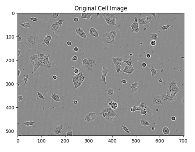
The initial step involves loading a VISTA-2D model checkpoint and using it to segment cells in an image. The segmentation process generates a feature vector for each cell, which contains all necessary information for cell morphology analysis. These vectors are then used in clustering algorithms to group cells with similar features.
Segmenting Cells
The segmentation function processes the cell image through VISTA-2D, resulting in segmentation masks that label each cell individually. This allows for accurate feature extraction for each cell.
img_path="example_livecell_image.tif"
patch, segmentation, pred_mask = segment_cells(img_path, model_ckpt)
Plotting Segmentation
The segmented images can be visually verified using the plot_segmentation function. This function displays the original image, the segmentation result, and individual masks for each cell.
plot_segmentation(patch, segmentation, pred_mask)


Clustering Features with RAPIDS
Once feature vectors are extracted, they are clustered using RAPIDS, a GPU-accelerated machine learning library. The TruncatedSVD algorithm reduces the dimensionality of the feature vectors, making it easier to visualize clusters in 3D space.
dim_red_model = TruncatedSVD(n_components=3)
X = dim_red_model.fit_transform(cell_features)
The DBSCAN algorithm is then used to cluster the reduced feature vectors. This method assigns cluster labels to each cell, which can be visualized using Plotly for an interactive 3D plot.
model = DBSCAN(eps=0.003, min_samples=2)
labels = model.fit_predict(X)

Conclusion
NVIDIA’s VISTA-2D model offers a significant advancement in cell imaging and spatial omics by providing accurate cell segmentation and feature extraction. Coupled with RAPIDS for clustering, this model enables efficient classification of cell types, paving the way for more detailed and automated biological research.
Image source: Shutterstock
Credit: Source link


 Bitcoin
  Ethereum
  Tether
  Solana
  XRP
XRP  Dogecoin
  USDC
USDC  Cardano
  Lido Staked Ether
  Avalanche
  TRON
TRON  Toncoin
  Wrapped stETH
  Shiba Inu
  Stellar
  Wrapped Bitcoin
  Polkadot
  Chainlink
  Bitcoin Cash
  WETH
WETH  Sui
Sui  Pepe
Pepe  NEAR Protocol
  LEO Token
  Uniswap
  Litecoin
  Wrapped eETH
  Aptos
Aptos  Internet Computer
  Hedera
  USDS
USDS  Cronos
  Ethereum Classic
  POL (ex-MATIC)
  Render
  Bittensor
  Ethena USDe
  Artificial Superintelligence Alliance
  Arbitrum
  Celestia
  Filecoin
  Dai
Dai  Stacks
  WhiteBIT Coin
  Bonk
Bonk  OKB
OKB  Cosmos Hub
Comments are closed.